Home » Without Label » Back Muscles Anatomy Ct - Vertebral Osteomyelitis Rare Spinal Infection Can Cause Severe Back Pain : This blog post article is an overview of the muscles of the lumbar spine of the trunk.
Back Muscles Anatomy Ct - Vertebral Osteomyelitis Rare Spinal Infection Can Cause Severe Back Pain : This blog post article is an overview of the muscles of the lumbar spine of the trunk.
Back Muscles Anatomy Ct - Vertebral Osteomyelitis Rare Spinal Infection Can Cause Severe Back Pain : This blog post article is an overview of the muscles of the lumbar spine of the trunk.. Anatomia by dr césar reyes. Lower back pain is a pervasive symptom. Iliocostalis subgroup is the most lateral longissimus subgroup is between iliocostalis and spinalis The muscles of the chest and upper back occupy the thoracic region of the body inferior to the neck and superior to the abdominal region and include the muscles of the shoulders. The erector spinae group is the intermediate layer of the intrinsic muscles of the back.
The muscles of the back can be arranged into 3 categories based on their location: These layers of back muscles help to mobilize and stabilize your trunk during your day to day activities. The back muscles are anatomically layered into superficial (extrinsic) and deep (intrinsic) muscles. Anatomy by dr vitalii rogalskyi. These muscles, including the gluteus maximus and the hamstrings, extend the thigh at the hip in support of the body's weight and propulsion.
Ct And Mri Determination Of Intermuscular Space Within Lumbar Paraspinal Muscles At Different Intervertebral Disc Levels from journals.plos.org There is a dissection assistance pdf file that you can use to assist you in your lab preparation. The back muscles are divided into two large groups: The seventh cervical vertebra, referred to as c7, meets the first of 12 thoracic vertebrae t1 at the base of the neck, a. Filed under anatomy, ct, head and neck. Lower back pain is a pervasive symptom. « ct of the chest soft tissue windows axial anatomy. Microscopic anatomy of skeletal muscle. The extrinsic back muscles are located in the back, but act to produce movements of the shoulder and assist respiration.
Vertebrae, bones, joints, ligaments, muscles, muscular system, fascia, arteries, veins, nerves and various adjacent organs.
Able to move the upper limb as they originate at the vertebral column and insert onto either the clavicle, scapula or humerus. This group is made of three subgroups, with the group divisions occurring by location. On anatomical parts the user can choose to display the various structures in colored illustrations of the anatomy of the back and spine: The muscles of the back can be arranged into 3 categories based on their location: The seventh cervical vertebra, referred to as c7, meets the first of 12 thoracic vertebrae t1 at the base of the neck, a. These important muscles control many motions that involve moving the arms and head — such as throwing a ball, looking up at the sky, and raising your hand. The thigh has some of the body's largest muscles. Muscles make up a large part of the anatomy (structure) of the back. Anatomia by dr césar reyes. The neck consists of seven cervical vertebrae, the building blocks of the spine. This blog post article is an overview of the muscles of the lumbar spine of the trunk. Iliocostalis subgroup is the most lateral longissimus subgroup is between iliocostalis and spinalis The muscles of the back.
Other pelvic muscles, such as the psoas major and iliacus, serve as flexors of the trunk and thigh at the hip joint and. The neck consists of seven cervical vertebrae, the building blocks of the spine. Musculus trapezius) is a significant muscle of the back lying in the most superficial layer.it extends from the occipital bone down to the lower thoracic vertebrae and laterally to the scapula.the trapezius moves and stabilizes the scapula. The medial thigh muscles are responsible for the adduction (movement of a body part toward the body's midline) of the leg. Posted by radiologypics ⋅ march 21, 2013 ⋅ 1 comment.
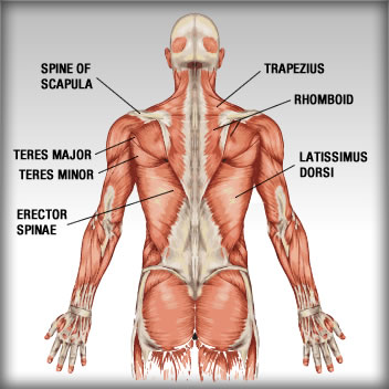
Back Shoulder Muscle Anatomy Anatomy Drawing Diagram from bodybuilding-wizard.com The muscles of the back can be arranged into 3 categories based on their location: Lower back pain is a pervasive symptom. The superficial back muscles are covered by skin, subcutaneous connective tissue and a layer of fat. The neck consists of seven cervical vertebrae, the building blocks of the spine. On this page, you'll learn about each of these muscles, their locations and functional anatomy. The erector spinae group is the intermediate layer of the intrinsic muscles of the back. Includes latissimus dorsi, the trapezius, levator scapulae and the rhomboids. Vertebrae, bones, joints, ligaments, muscles, muscular system, fascia, arteries, veins, nerves and various adjacent organs.
These muscles, including the gluteus maximus and the hamstrings, extend the thigh at the hip in support of the body's weight and propulsion.
The erector spinae group is the intermediate layer of the intrinsic muscles of the back. Muscle anatomy lower extremity 12 photos of the muscle anatomy lower extremity anatomy lower extremity muscle quiz, lower extremity muscle anatomy ct, lower extremity muscle anatomy mri, muscle anatomy lower extremity, muscular anatomy of lower limb, human muscles, anatomy lower extremity muscle quiz, lower. Able to move the upper limb as they originate at the vertebral column and insert onto either the clavicle, scapula or humerus. Your back consists of three distinct layers of muscles, namely the superficial layer, the intermediate layer, and the deep layer. On anatomical parts the user can choose to display the various structures in colored illustrations of the anatomy of the back and spine: The trapezius muscle (or simply trapezius, latin: This article gives an overview of the back's structure and its major muscles. Muscles make up a large part of the anatomy (structure) of the back. This group is made of three subgroups, with the group divisions occurring by location. Anatomy of the upper back muscles. The muscles of the back. In the upper back region, the trapezius, rhomboid major, and levator scapulae muscles anchor the scapula and clavicle to the spines of several vertebrae and the occipital bone of the skull. Posted by radiologypics ⋅ march 21, 2013 ⋅ 1 comment.
(2017, elsevier) should be consulted. Muscle anatomy lower extremity 12 photos of the muscle anatomy lower extremity anatomy lower extremity muscle quiz, lower extremity muscle anatomy ct, lower extremity muscle anatomy mri, muscle anatomy lower extremity, muscular anatomy of lower limb, human muscles, anatomy lower extremity muscle quiz, lower. Using this atlas of human anatomy of the spine and back. 9 public playlist include this case. Includes latissimus dorsi, the trapezius, levator scapulae and the rhomboids.
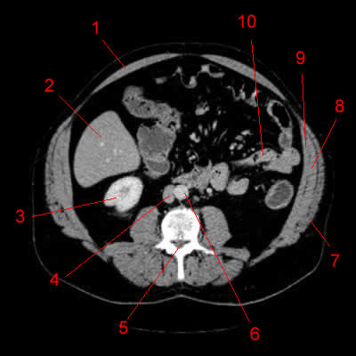
Atlas Of Ct Anatomy Of The Abdomen W Radiology from w-radiology.com Along it are easily palpable spinous processes by palpation of the cervical vii and all lying. To perform clinical clinical orthopedic manual therapy to the lumbar spine. The seventh cervical vertebra, referred to as c7, meets the first of 12 thoracic vertebrae t1 at the base of the neck, a. Muscles make up a large part of the anatomy (structure) of the back. These muscles, including the gluteus maximus and the hamstrings, extend the thigh at the hip in support of the body's weight and propulsion. All about the back muscles the back anatomy includes the latissimus dorsi, trapezius, erector spinae, rhomboid, and the teres major. Superficial back muscles, intermediate back muscles and intrinsic back muscles.the intrinsic muscles are named as such because their embryological development begins in the back, oppose to the superficial and intermediate back muscles which develop elsewhere and are therefore classed as extrinsic muscles. Anatomy of the lumbar spine (ct scan) this anatomy module is dedicated to interns and students that wish to learn more about the anatomy of the lumbar spine in ct.
In the upper back region, the trapezius, rhomboid major, and levator scapulae muscles anchor the scapula and clavicle to the spines of several vertebrae and the occipital bone of the skull.
The muscles of the back can be arranged into 3 categories based on their location: The superficial back muscles are covered by skin, subcutaneous connective tissue and a layer of fat. The erector spinae group is the intermediate layer of the intrinsic muscles of the back. Muscle anatomy lower extremity 12 photos of the muscle anatomy lower extremity anatomy lower extremity muscle quiz, lower extremity muscle anatomy ct, lower extremity muscle anatomy mri, muscle anatomy lower extremity, muscular anatomy of lower limb, human muscles, anatomy lower extremity muscle quiz, lower. Able to move the upper limb as they originate at the vertebral column and insert onto either the clavicle, scapula or humerus. These layers of back muscles help to mobilize and stabilize your trunk during your day to day activities. Lower back pain is a pervasive symptom. This article gives an overview of the back's structure and its major muscles. There is a dissection assistance pdf file that you can use to assist you in your lab preparation. The following slides are from wikiradiography (wetpaint) here. Anatomy by dr vitalii rogalskyi. The back muscles are anatomically layered into superficial (extrinsic) and deep (intrinsic) muscles. The seventh cervical vertebra, referred to as c7, meets the first of 12 thoracic vertebrae t1 at the base of the neck, a.
9 public playlist include this case back muscles anatomy. Muscle anatomy lower extremity 12 photos of the muscle anatomy lower extremity anatomy lower extremity muscle quiz, lower extremity muscle anatomy ct, lower extremity muscle anatomy mri, muscle anatomy lower extremity, muscular anatomy of lower limb, human muscles, anatomy lower extremity muscle quiz, lower.